by Ruth Beran
X-ray imaging is widely used to record what is inside objects. X-rays are taken in hospitals to look at bone fractures, at dentists to look for cavities in teeth, and by vets to look at breaks in race horses' legs. X-rays are also used by engineers to look for cracks in pipelines and at the airport to image the contents of suitcases during security checks.
At Victoria University of Wellington, a portable X-ray detector has been developed which could allow people like medical responders to take X-ray images on the scene in an emergency and send them to hospitals or treatment centres before a patient even arrives.
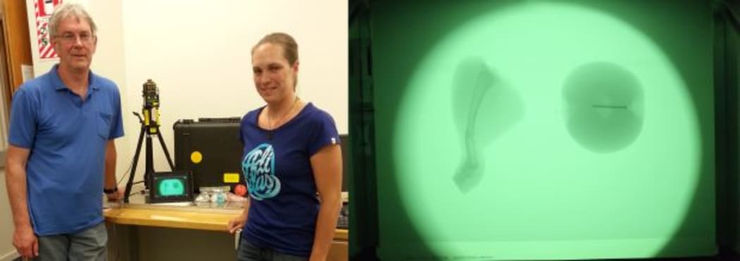
From left to right: Andy Edgar and Nicola Winch with the portable X-ray detector, and an image of a broken chicken leg and apple with a nail in it Photo: RNZ / VUW
The system was developed by PhD student Nicola Winch to test materials for recording X-ray images created by supervisor Andy Edgar and his team, and consists of two components, the read out unit contained in a hard-shelled box and a commercial x-ray generator which sits on a tripod. The imaging plate is in the box, which can be removed. The box is of a size that would take a laptop or a musical instrument. The system “uses an optical technique, so using light to read the x-ray images,” says Winch “The box is very robust and can withstand the knocks and is also light tight since we’re using optical methods.”
The detector uses a large number of custom made items put together in an integrated fashion. “One of the neat features about this one is we can use smart phones to take these images, or iPads or laptops, so it’s totally wireless and it will connect via standalone wireless networks,” says Winch. It’s also light and rugged. “So you could take it just about anywhere you wanted,” she says. The image can be transmitted easily over a range of 20 or 30 metres and the image recorded can reach a size of 20MB, but would be limited to the resolution of the receiving device.
The system offers portability, is light weight, has very easy communications and ease of use. “We see it having great potential in many applications,” says Edgar. “Of course our original purpose was to demonstrate how our own imaging plates and materials work, and still that’s its immediate use for us, but we can see it having much wider applications than just that.”
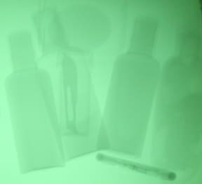
X-ray image of toiletries with hidden scalpel Photo: VUW
Edgar’s team are also developing materials for recording x-ray images, to replace photographic film which has been used for at least 100 years. These materials are used to record the passage of x-rays through materials to highlight dense objects inside lighter ones. “For example, bones in tissue stand out of course in an x-ray image,” says Edgar.
The materials being developed are sheets of glass. “When I talk of a glass people think of window glass or the glass they have in spectacles,” says Edgar. “Those are long standing historic glasses, but the term glass doesn’t really refer to a specific chemical composition…it’s a state of matter. It’s a random organisation of the atoms in a solid material.”
The glasses Edgar and his team work on are very different to those used in window glass, instead of silica or phosphorous oxides they are based on fluoride. These materials are made in argon gas to prevent a reaction with the water vapour in air. They are melted in a radio frequency induction furnace, where the power of a radio transmitter is concentrated into a small crucible containing the glass material which is melted, poured onto a cold plate and frozen into its amorphous state.
The glass looks transparent, but is doped. So if ultraviolet light is shone onto it, it glows red. If it is X-rayed, it glows orange. “So if we pick out the orange light and discard the red light we have a direct image of where X-rays have fallen on the plate and hence an image of whatever was placed in the X-ray beam between the plate and the source of X-rays,” says Edgar.
These materials record with greater sensitivity and require fewer X-rays to develop the image, which is better for people’s health. “You can record an image and then erase the image and use the same imaging plate,” says Edgar. “We make a plate out of this material, use the same plate time and time again, just by simply erasing it each time you use it, there’s no nasty chemicals involved which is for example in photographic film where one has to use developer and so forth.” They are also compatible with computers.