By Ruth Beran
Each day approximately 17 New Zealanders – one every 90 minutes – die as a result of coronary heart disease.
People suffering from coronary heart disease have a narrowing or blockage of their coronary arteries by plaque formed by fat or cholesterol on the artery walls.
The preferred treatment is to insert a stent, and PhD student Susann Beier is trying to improve stent design.
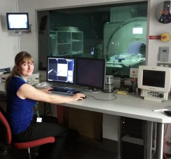
Susann Beier in the MRI control room Photo: RNZ / Ruth Beran
A stent is a tiny meshed wire tube which has been put in the circulatory system to be entered into the narrowed arteries,” says Susann.
Stents are used to recover blood flow in an artery and they are deployed in arteries with the help of a balloon. “[The balloon] compresses the arterial plug against the vessel wall which opens the vessel up again,” says Susann.
Stents are usually inserted around the heart and this is the area where Susann Beier is focusing her research. However, stents can also be used in the carotid arteries for stroke prevention.
In particular, Susann is looking at blood flow and stented coronaries. Her aim is to improve stent design and identify predictors for coronary artery disease.
She is using two powerful tools in her research: computational modelling of stents and MRI (magnetic resonance imaging) to measure blood flow in large replicas of stented arteries.
The replicas are 3D printed, are six times the size of a human heart, and look like a piped “Y” depicting one coronary branch when it divides into two.
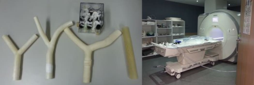
From the left to right: 3D-printed replicas of coronary arteries and the flow setup in the MRI Photo: RNZ / Ruth Beran
Just like humans look very different our arteries look very different as well,” says Susann.
For example, one replica has a very wide branching artery with 110 degrees in between the daughter branches. Another branches only by 30 degrees.
To measure the blood flow in the up-scaled artery replica, a flow circuit pumps blood-like fluid through and it is then placed in an MRI scanner. The MRI then measures flow in 4D, i.e. in three dimensions at each point in time. About 100 litres of blood like fluid is pumped along pipelines using equipment which must be located outside the MRI because nothing magnetic can be taken into the room.
Susann then compares the outputs from her real-scale computer simulation with the measurements from the MRI to see how closely they correlate. “This is the first time anybody has ever done this and they correlate really well,” she says.
By measuring flow, Susann can see where there is really low flow or high flow in the replicas. This in turn puts low or high stress on the arteries. “Then we can say disease is likely to grow there,” she says.
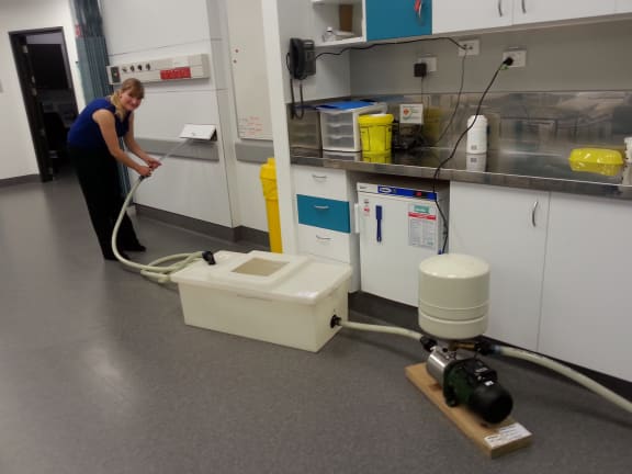
Susann Beier with the pipes and flow pump for the bloodlike fluid outside the MRI room Photo: RNZ / Ruth Beran
The fluid is pumped from the single inlet at the bottom of the “Y” and then it divides into the two daughter branches. “At times it creates swirls and stagnation zones. These are the things we’re looking for and then it goes back into the single pipe and comes back around,” she says.
Susann has many replicas, with stents and without. She also has idealised replicas, ones that are average, and some patient specific ones.
Ultimately, she hopes that her work will lead to improved stent design.
“If we implant a stent into a vessel in most cases it’s really helpful and it’s great,” says Susann.
However, in a very small percentage of cases the design of the stent actually changes the flow environment in a way which is not beneficial.
So if we can identify those specific stent design features then we can optimise stent design. And improve stent success,” she says.
Susann conducts her work at the Centre for Advanced MRI at the University of Auckland. CAMRI is involved in a lot of clinical studies but also allows PhD students like Susann to conduct research there.